


ARTICLE CONTENTSOn this page 11 sections
Cell Structure and Functions
Performance Objectives
By the end of the lesson, students should be able to:
- identify parts of the cell under the microscope;
- describe the structure of parts of the cell using appropriate biological terms;
- match each organelle with its specific function in the cell;
- draw and label plant and animal cells;
- differentiate between plant and animal cells;
- describe the stages of mitosis and meiosis with diagrams;
- demonstrate understanding of diffusion and osmosis through simple experiments or simulations.
Introduction to Cells
The cell is the basic structural and functional unit of all known living organisms. It is the smallest unit of life that can replicate independently. All living things, from the smallest bacteria to the largest trees and animals, are made up of cells.
Cells are often called the “building blocks of life” because they perform all the essential processes necessary for an organism to survive, grow, and reproduce. Despite their small size, cells are highly organised and contain many specialised structures that carry out specific tasks.
General Cell Structure
All cells share some common features. They are enclosed by a cell membrane, which separates the inside of the cell from its external environment. Inside the cell membrane is the cytoplasm, a jelly-like substance that fills the cell and contains various organelles. Organelles are like tiny organs, each with a specific role to play in the cell’s overall function.
Animal Cell Structure
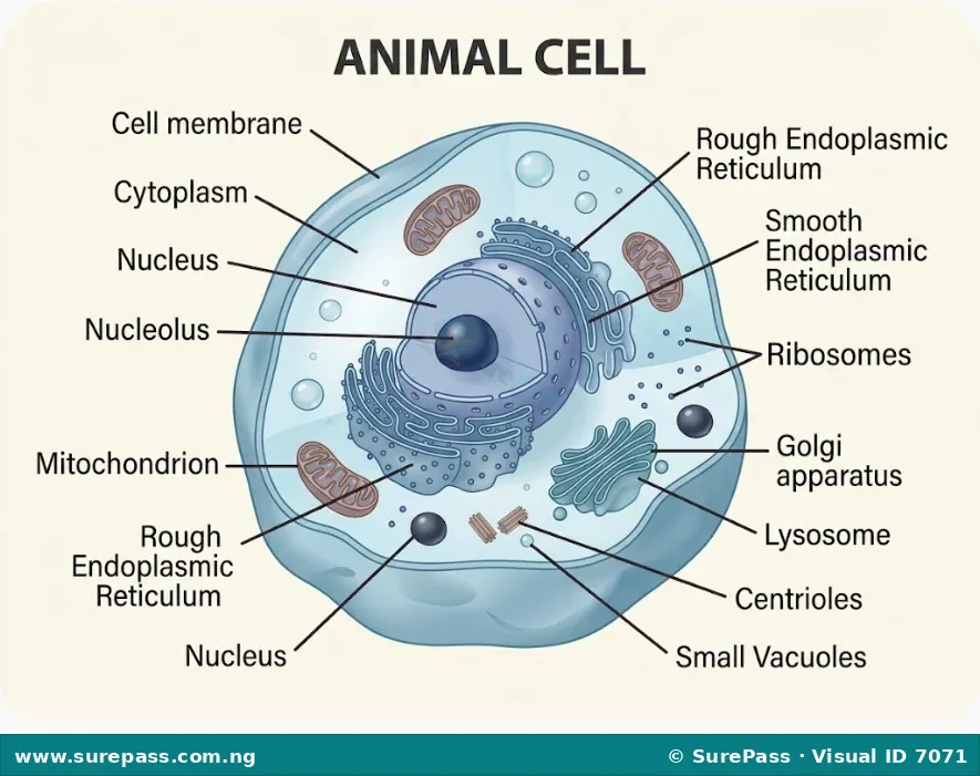
Animal cells are typically round or irregular in shape. They are eukaryotic, meaning they have a true nucleus and other membrane-bound organelles. Key components of an animal cell include the cell membrane, cytoplasm, nucleus, mitochondria, endoplasmic reticulum, ribosomes, Golgi apparatus, lysosomes, and small vacuoles.
Plant Cell Structure
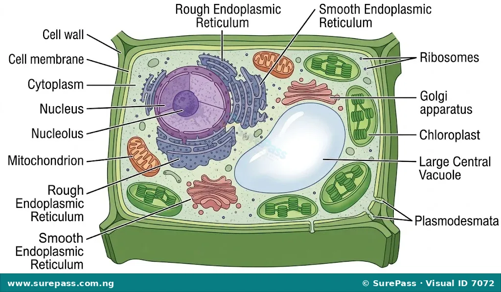
Plant cells are typically rectangular or fixed in shape due to the presence of a rigid cell wall outside the cell membrane. Like animal cells, they are eukaryotic. In addition to the organelles found in animal cells, plant cells have a cell wall, chloroplasts, and a large central vacuole. These additional structures are crucial for photosynthesis and maintaining cell shape.
Functions of Cell Organelles
Each organelle within a cell performs a specific function vital for the cell’s survival and proper operation. Understanding these functions helps to appreciate the complexity and efficiency of cellular life.
-
Cell Membrane:
The cell membrane is a thin, flexible outer boundary of the cell. It controls the movement of substances into and out of the cell, acting as a selective barrier. It is made up of a double layer of lipids and proteins.
-
Cytoplasm:
The cytoplasm is the jelly-like substance that fills the cell, surrounding all the organelles. It is the site where many metabolic reactions and cellular processes occur, such as glycolysis.
-
Nucleus:
The nucleus is a large, spherical organelle that contains the cell’s genetic material (DNA) organised into chromosomes. It controls all cellular activities, including growth, metabolism, and reproduction. The nucleus is often called the “control centre” of the cell.
-
Mitochondria:
Mitochondria are often referred to as the “powerhouses” of the cell. They are responsible for cellular respiration, a process that converts glucose and oxygen into energy (ATP) for the cell’s activities.
-
Endoplasmic Reticulum (ER):
The endoplasmic reticulum is a network of membranes found throughout the cytoplasm of eukaryotic cells. It plays a key role in the synthesis, folding, modification, and transport of proteins and lipids. There are two types: rough ER (with ribosomes) and smooth ER (without ribosomes).
-
Ribosomes:
Ribosomes are small organelles responsible for protein synthesis. They can be found floating freely in the cytoplasm or attached to the rough endoplasmic reticulum.
-
Golgi Apparatus (or Golgi Complex/Body):
The Golgi apparatus modifies, sorts, and packages proteins and lipids into vesicles for secretion or delivery to other organelles. It is like the cell’s postal service.
-
Lysosomes:
Lysosomes are membrane-bound sacs containing digestive enzymes. They break down waste materials, cellular debris, and foreign invaders like bacteria, acting as the cell’s recycling and waste disposal units.
-
Vacuoles:
Vacuoles are membrane-bound sacs that store water, nutrients, and waste products. In plant cells, a large central vacuole helps maintain turgor pressure, supporting the cell’s shape. Animal cells have smaller, temporary vacuoles.
-
Cell Wall (in plant cells):
The cell wall is a rigid outer layer found only in plant cells, outside the cell membrane. It provides structural support, protection, and prevents excessive water uptake. It is primarily made of cellulose.
-
Chloroplasts (in plant cells):
Chloroplasts are organelles found only in plant cells and some algae. They are the sites of photosynthesis, the process by which plants convert light energy into chemical energy (glucose) using chlorophyll.
-
Centrioles (in animal cells):
Centrioles are cylindrical structures found in animal cells, usually near the nucleus. They are involved in cell division, helping to organise the spindle fibres that separate chromosomes.
Differences between Plant and Animal Cells
While both plant and animal cells are eukaryotic and share many common organelles, they also have distinct differences that reflect their different functions and lifestyles.
-
Cell Wall: Plant cells have a rigid cell wall made of cellulose, which provides structural support and protection. Animal cells lack a cell wall.
-
Chloroplasts: Plant cells contain chloroplasts, which are responsible for photosynthesis due to the presence of chlorophyll. Animal cells do not have chloroplasts or chlorophyll.
-
Vacuoles: Plant cells typically have one large, permanent central vacuole that can occupy up to 90% of the cell volume, maintaining turgor pressure. Animal cells usually have several small, temporary vacuoles, or none at all.
-
Shape: Due to the cell wall, plant cells have a fixed, often rectangular or cuboid shape. Animal cells have an irregular or round shape, as they lack a rigid cell wall.
-
Centrioles: Animal cells possess centrioles, which are involved in cell division. Plant cells generally lack centrioles.
-
Food Storage: Plant cells store food primarily as starch. Animal cells store food as glycogen.
The table below summarises the key differences between plant and animal cells:
| Feature | Plant Cell | Animal Cell |
|---|---|---|
| Cell Wall | Present (made of cellulose) | Absent |
| Chloroplasts | Present | Absent |
| Vacuole | One large central vacuole | Small, temporary, or absent |
| Shape | Fixed, often rectangular | Irregular or round |
| Centrioles | Absent (except in lower plants) | Present |
| Food Storage | Starch | Glycogen |
Cell Division
Cell division is the process by which a parent cell divides into two or more daughter cells. It is essential for growth, repair, and reproduction in living organisms. The two main types of cell division are mitosis and meiosis.
Mitosis
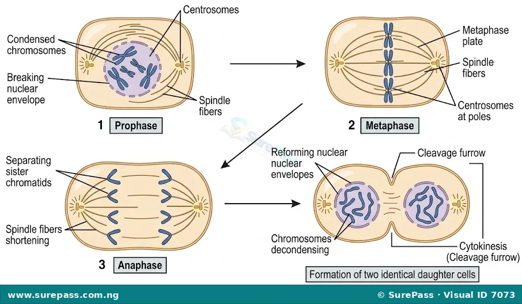
Mitosis is a type of cell division that results in two daughter cells each having the same number and kind of chromosomes as the parent nucleus. This process is essential for growth, repair of damaged tissues, and asexual reproduction in some organisms.
The stages of mitosis are:
-
Prophase: Chromosomes condense and become visible. The nuclear envelope begins to break down, and the spindle fibres start to form.
-
Metaphase: Chromosomes align themselves along the metaphase plate (equator) of the cell. Each chromosome is attached to a spindle fibre at its centromere.
-
Anaphase: Sister chromatids (identical halves of a chromosome) separate and move to opposite poles of the cell, pulled by the spindle fibres.
-
Telophase: Chromosomes arrive at the poles and begin to decondense. New nuclear envelopes form around the two sets of chromosomes. The spindle fibres disappear, and cytokinesis (division of the cytoplasm) usually begins.
Meiosis
Meiosis is a specialised type of cell division that reduces the chromosome number by half, creating four haploid cells, each genetically distinct from the parent cell. This process is essential for sexual reproduction, as it produces gametes (sperm and egg cells) in animals and spores in plants.
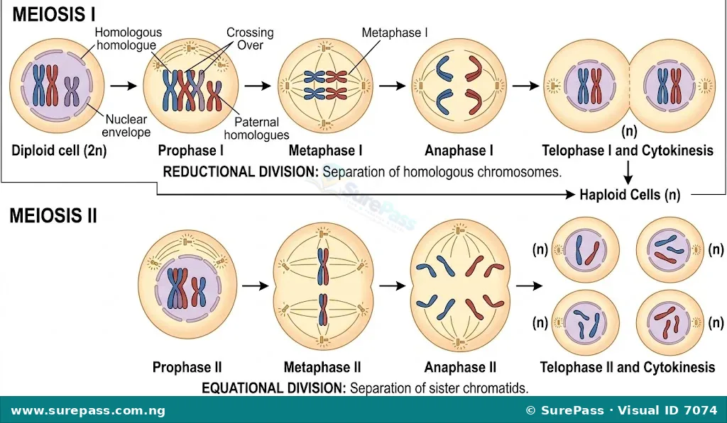
Meiosis involves two rounds of division: Meiosis I and Meiosis II.
Meiosis I:
-
Prophase I: Chromosomes condense, homologous chromosomes pair up (forming bivalents), and crossing over (exchange of genetic material) occurs. The nuclear envelope breaks down.
-
Metaphase I: Homologous chromosome pairs align at the metaphase plate.
-
Anaphase I: Homologous chromosomes separate and move to opposite poles of the cell. Sister chromatids remain attached.
-
Telophase I: Chromosomes arrive at the poles, and new nuclear envelopes may form. Cytokinesis usually follows, resulting in two haploid daughter cells, each with duplicated chromosomes.
Meiosis II:
Meiosis II is similar to mitosis, but it occurs in the two haploid cells produced during Meiosis I.
-
Prophase II: Chromosomes condense again, and the nuclear envelope breaks down (if it reformed).
-
Metaphase II: Sister chromatids align at the metaphase plate of each cell.
-
Anaphase II: Sister chromatids separate and move to opposite poles.
-
Telophase II: Chromosomes arrive at the poles, new nuclear envelopes form, and cytokinesis occurs. This results in four haploid daughter cells, each with unduplicated chromosomes.
Cell Processes
Cells are constantly interacting with their environment, exchanging substances to maintain their internal balance. Key processes involved in this exchange include diffusion, osmosis, plasmolysis, and haemolysis.
Diffusion
Diffusion is the net movement of particles from a region of higher concentration to a region of lower concentration. This movement occurs down a concentration gradient until the particles are evenly distributed. Diffusion does not require energy and can occur in gases, liquids, and even solids.
Examples of diffusion in living systems include the movement of oxygen from the lungs into the bloodstream, the movement of carbon dioxide from the bloodstream into the lungs, and the absorption of digested food molecules from the small intestine into the blood.
Osmosis
Osmosis is the net movement of water molecules across a selectively permeable membrane from a region of higher water concentration (lower solute concentration) to a region of lower water concentration (higher solute concentration). Like diffusion, osmosis does not require energy.
A selectively permeable membrane allows water molecules to pass through but restricts the movement of larger solute molecules. This process is crucial for water balance in cells and organisms.
Plasmolysis
Plasmolysis is the process in plant cells where the protoplast (cell membrane and its contents) shrinks and pulls away from the cell wall when the cell is placed in a hypertonic solution (a solution with a higher solute concentration than the cell’s cytoplasm). Water moves out of the cell by osmosis, causing the cell to lose turgor pressure and become flaccid.
If a plant cell remains plasmolyzed for too long, it can die. This process demonstrates the importance of water balance for plant cells.
Haemolysis
Haemolysis is the bursting of red blood cells when they are placed in a hypotonic solution (a solution with a lower solute concentration than the cell’s cytoplasm). In this situation, water moves into the red blood cells by osmosis, causing them to swell and eventually burst because they lack a rigid cell wall to withstand the increased internal pressure.
This process highlights the fragility of animal cells in solutions with very low solute concentrations.
Observing Cells Under the Microscope
Observing cells under a microscope allows for direct identification of their parts and comparison between different cell types. This skill is fundamental in biology.
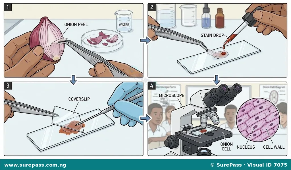
Procedure for Preparing and Observing an Onion Epidermal Cell
The onion epidermal cell is a common example of a plant cell used for microscopic observation due to its large, easily visible cells.
The required materials are:
- Onion bulb
- Spatula or forceps
- Microscope slide
- Coverslip
- Dropper
- Water
- Methylene blue stain (optional, but helps visibility)
- Light microscope
The procedure involves the following steps:
- Carefully peel off a thin, transparent layer (epidermis) from the inner surface of an onion scale leaf using a spatula or forceps. Try to get a single layer.
- Place a drop of water in the centre of a clean microscope slide. If using a stain, add a small drop of methylene blue to the water.
- Gently place the onion epidermis flat in the water/stain drop on the slide. Ensure there are no folds or air bubbles.
- Carefully lower a coverslip over the specimen at an angle to avoid trapping air bubbles.
- Place the slide on the microscope stage. Start with the lowest power objective lens (e.g., 4x or 10x).
- Adjust the coarse adjustment knob to bring the specimen into rough focus, then use the fine adjustment knob for clear focus.
- Observe the cells. Identify the cell wall, cell membrane, cytoplasm, and nucleus. The large central vacuole may be visible as a clear area.
- Switch to a higher power objective lens (e.g., 40x) for a more detailed view, adjusting the fine adjustment knob as needed.
Drawing Cell Structure
Drawing biological specimens accurately is an important skill that helps in understanding and remembering structures. When drawing cells, certain conventions should be followed.
Guidelines for drawing cell structures:
-
Use a sharp pencil: Drawings should be clear and neat.
-
Draw large: Make the drawing large enough to show details clearly, occupying a good portion of the available space.
-
Use continuous lines: Avoid sketchy or broken lines. Draw smooth, continuous outlines.
-
Label clearly: Use straight, uncrossed label lines that point directly to the structure being labelled. Write labels horizontally.
-
Include a title: Every drawing should have a clear, descriptive title (e.g., “Diagram of an Animal Cell”).
-
Indicate magnification: If drawn from a microscope, state the magnification used (e.g., “x400”).
-
Do not shade: Biological drawings are typically outlines, not shaded. Stippling (dots) can be used to indicate density if necessary, but shading is generally avoided.
Demonstrating Osmosis and Diffusion
Simple experiments can help demonstrate the principles of osmosis and diffusion, making these abstract concepts more concrete.
Demonstrating Diffusion
Diffusion can be observed by watching the spread of a coloured substance in water.
The required materials are:
- Beaker or clear glass
- Water
- Potassium permanganate crystals or a drop of food colouring
The procedure involves the following steps:
- Fill the beaker with clear water.
- Carefully drop a few crystals of potassium permanganate or a drop of food colouring into the water without stirring.
- Observe the beaker over time.
- The colour will slowly spread throughout the water, moving from a region of high concentration (where the crystals/drop were placed) to a region of lower concentration until it is evenly distributed. This demonstrates diffusion.
Demonstrating Osmosis using an Onion Cell
The effect of osmosis on plant cells can be observed using onion epidermal cells and solutions of different concentrations.
The required materials are:
- Onion bulb
- Spatula or forceps
- Microscope slides
- Coverslips
- Dropper
- Distilled water (hypotonic solution)
- Concentrated salt solution (hypertonic solution)
- Light microscope
The procedure involves the following steps:
- Prepare two separate slides with onion epidermal cells, following steps 1-4 from the “Observing Cells Under the Microscope” section.
- For the first slide, use distilled water as the mounting medium. This represents a hypotonic environment.
- For the second slide, use a concentrated salt solution as the mounting medium. This represents a hypertonic environment.
- Observe both slides under the microscope, starting with low power and moving to high power.
- On the slide with distilled water, the onion cells will appear turgid (swollen) and pressed against the cell wall. This is because water moves into the cells by osmosis.
- On the slide with salt solution, the onion cells will show plasmolysis. The protoplast (cell membrane and its contents) will shrink and pull away from the rigid cell wall, leaving a gap. This is because water moves out of the cells by osmosis.
- Compare the appearance of the cells in both solutions to understand the effect of osmosis.




0 comments
Open the discussion when you need it. Comments stay collapsed until requested so the initial article remains light.